What the study found
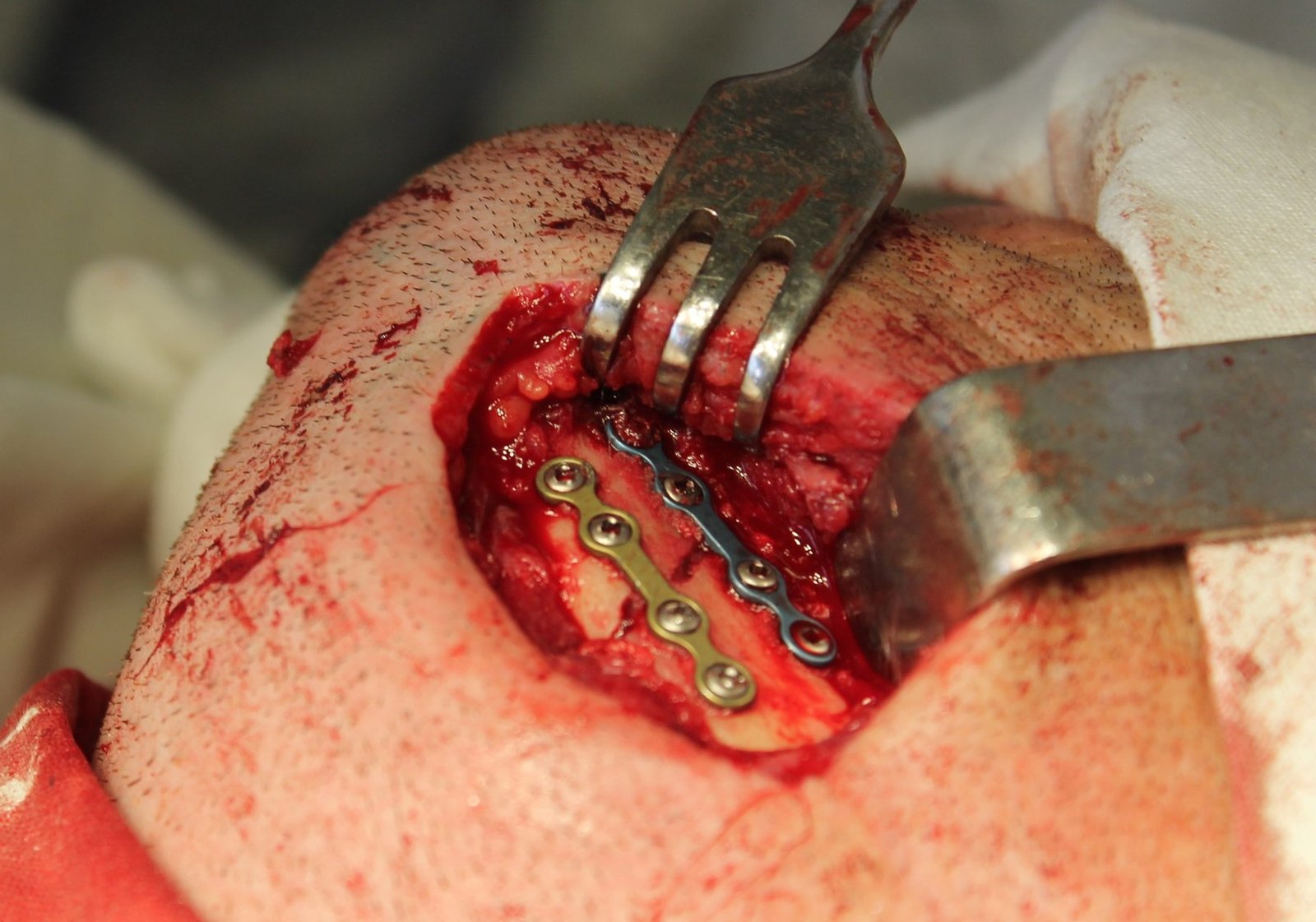
The study found that titanium osteosynthesis plate design affected both how radiation was distributed near the plates and how two human cell types grew after exposure. In particular, plate geometry and diameter were associated with dose increases in front of the plates and dose reductions behind them.
Why the authors say this matters
The authors conclude that these findings matter because metallic fixation hardware may influence dose distribution during adjuvant radiotherapy, which is radiotherapy given after surgery. They suggest that reduced metal volume and specific plate geometries should be considered in reconstructive planning for patients undergoing this treatment.
What the researchers tested
The researchers used a three-dimensional printed mandibular phantom based on patient CT data, together with a water phantom, to simulate clinical irradiation conditions. They irradiated the setup with an Ethos linear accelerator and analyzed dose changes near different plate designs. They also irradiated human mesenchymal stromal cells and human umbilical vein endothelial cells under the same conditions and measured proliferation with MTS assays.
What worked and what didn't
Distinct dose increases were detected anterior to the plates, and dose reductions were seen in the shadowing region. These physical changes were associated with significant differences in proliferation for both human mesenchymal stromal cells and human umbilical vein endothelial cells. The effects varied depending on plate diameter and geometry.
What to keep in mind
This was an experimental study using a printed phantom and cell culture models, so the summary does not provide direct clinical outcome data from patients. The abstract does not describe additional limitations beyond the experimental setup.
Key points
- Titanium osteosynthesis plate geometry changed local radiation dose patterns.
- Dose increases occurred in front of the plates, with dose reductions behind them.
- Cell proliferation of human mesenchymal stromal cells and human umbilical vein endothelial cells differed after irradiation.
- The effects depended on plate diameter and geometry.
- The authors suggest considering reduced metal volume and specific plate shapes in reconstructive planning.
Disclosure
- Research title:
- Plate design altered radiation dose and cell growth
- Authors:
- Georg Hoene, Andre Tangemann, Boris Schminke, Lennart Johannes Gruber, Martin Leu, Daniela Schmitt, Henning Schliephake, Philipp Kauffmann
- Institutions:
- Universitätsmedizin Göttingen, Universitätsmedizin Göttingen, Universitätsmedizin Göttingen, Universitätsmedizin Göttingen, Universitätsmedizin Göttingen, Universitätsmedizin Göttingen, Universitätsmedizin Göttingen, University of Göttingen
- Publication date:
- 2026-02-25
- OpenAlex record:
- View
- Image credit:
- Katia&Lena, Wikimedia Commons, CC BY-SA 4.0
Get the weekly research newsletter
Stay current with scholarly research without reading academic papers — one filtered digest, every Friday.