Tag: Diagnostics & Medical Imaging

  • Favorable second molar eruption signs were uncommon at first molar extraction

    What the study found

    The study found that radiographic signs associated with favorable spontaneous eruption of the second permanent molar were present in only a limited share of children when first permanent molar extraction was being considered. The authors report that this gap reflects a mismatch between ideal developmental timing and the dental disease that often leads to extraction.

    Why the authors say this matters

    The authors conclude that systematic radiographic evaluation of second permanent molar developmental stage, angulation, and third permanent molar germ presence should be part of individualized treatment planning. The study suggests this is important because extraction is often required after advanced structural disease has already developed.

    What the researchers tested

    This retrospective study reviewed panoramic radiographs from 500 pediatric patients with extraction-indicated first permanent molars, involving 712 teeth. The researchers recorded demographic features, dentition stage, extraction indications, second permanent molar developmental stage using the Demirjian classification, second permanent molar angulation, and the presence of a third permanent molar germ.

    What worked and what didn't

    Extensive carious destruction was the most common extraction indication, followed by repeated treatment interventions, severe structural breakdown, and molar-incisor hypomineralization-related defects. Among the radiographic findings, 17.6% of second permanent molars were at Demirjian stage E, 27.4% were at stage F, 55.2% showed mesial angulation, and 74.2% had a third permanent molar germ present. Only 12.8% of cases met all predefined radiographic conditions associated with favorable eruption potential after first permanent molar extraction.

    What to keep in mind

    The study is retrospective and based on panoramic radiographs from one pediatric sample, so it describes associations rather than testing a treatment effect. The abstract does not describe longer-term clinical follow-up of eruption outcomes for all cases, and it does not report limitations beyond the restricted availability of favorable radiographic conditions.

    • Only 12.8% of cases met all predefined radiographic conditions linked to favorable second permanent molar eruption.
    • Extensive carious destruction was the main reason for first permanent molar extraction.
    • Most second permanent molars were not at the most favorable developmental stage, and many showed mesial angulation.
    • A third permanent molar germ was present in 74.2% of cases.
    • The authors recommend systematic radiographic assessment during individualized treatment planning.

  • Deep learning quantified craniofacial growth and sex differences

    What the study found

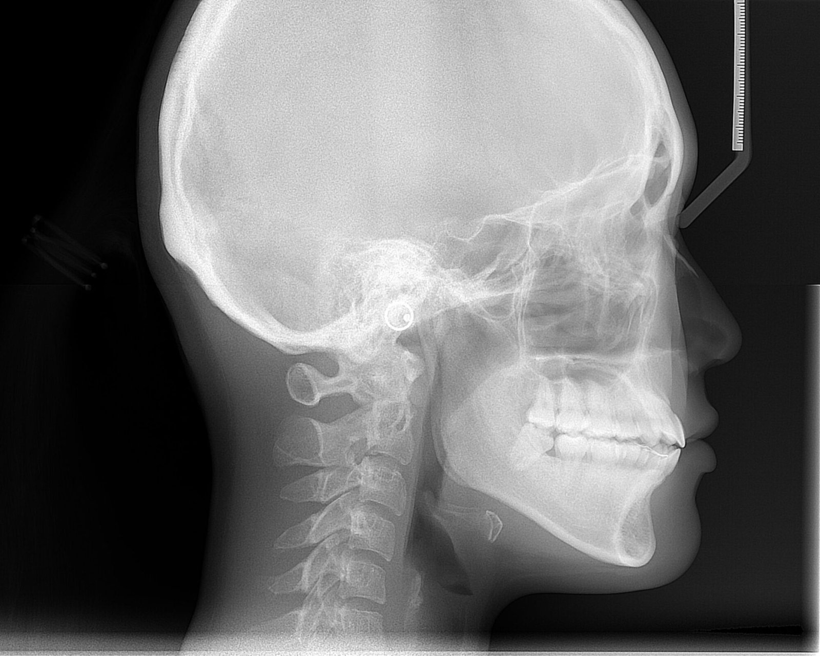
    The study found that an end-to-end deep learning framework could analyze craniofacial growth across childhood and adolescence using lateral cephalometric radiographs. It also identified age-related and sex-related patterns in craniofacial skeletal regions and quantified them with new indices.

    Why the authors say this matters

    The authors conclude that the findings provide objective quantitative references for assessing developmental stages and guiding the timing of interventions targeting specific craniofacial regions. They also say the results validate established developmental theories and offer new insight into coordinated bone growth and sex-specific radiological characteristics.

    What the researchers tested

    The researchers developed an end-to-end deep learning framework using lateral cephalometric radiographs from 41,625 people aged 4–18 years. The model was designed to extract features linked to continuous age intervals and sexual dimorphism without manual annotations, and Gradient-weighted Class Activation Mapping (Grad-CAM), a method for visualizing model attention, was used to generate population-averaged saliency maps. They also introduced two quantitative measures: the Age-related Saliency Index (ASI) and the Sex-related Saliency Index (SSI).

    What worked and what didn't

    Age-related saliency maps extended the focus from external contours to internal anatomical details of the bones. The ASI was used to prioritize regions by age-related importance, and the SSI showed that early sex differences were widely distributed across cranial bones but became concentrated in the mandibular region by adulthood. The abstract does not report specific failed approaches or negative results.

    What to keep in mind

    The summary does not describe detailed limitations, comparison models, or performance metrics. The findings are based on lateral cephalometric radiographs from ages 4–18, so the stated scope is limited to that population and imaging type.

    • The study used 41,625 lateral cephalometric radiographs from people aged 4–18 years.
    • An end-to-end deep learning framework was built without manual annotations.
    • Grad-CAM was used to visualize age-related and sex-related model features.
    • Age-related maps highlighted internal bone details as well as external contours.
    • Sex-related differences were described as broad early on and later concentrated in the mandible.
    • The authors say the results may help assess developmental stage and intervention timing.
  • Care shapes anatomical pathology technologists’ autopsy practice

    Care shapes anatomical pathology technologists’ autopsy practice

    What the study found

    The study found that care is woven throughout the practice and identities of Anatomical Pathology Technologists, who assist during post-mortems and care for the deceased body before and after autopsy. The author argues that this care can be both technically and morally good, including actions that go beyond what is necessary or required.

    Why the authors say this matters

    The authors suggest this helps extend understandings of care to relationships with the dead. They also conclude that it adds new insight into how coronial justice can, and should, gain legitimacy.

    What the researchers tested

    The article uses original empirical data from interviews with Anatomical Pathology Technologists. These are professionals who assist in medico-legal autopsies and take responsibility for the deceased person's body before and after the post-mortem examination.

    What worked and what didn't

    The interviews support the argument that care is present throughout this work, even within a setting shaped by complex relations and regulations. The abstract says this care can include actions that go beyond what is necessary or mandated.

    What to keep in mind

    The abstract does not describe specific limitations, sample size, or the interview design in detail. The findings are based on Anatomical Pathology Technologists' accounts in the context of medico-legal autopsies.

    • The study says care is central to Anatomical Pathology Technologists' work in medico-legal autopsies.
    • The author argues that care can be technically and morally good, including actions beyond formal requirements.
    • The article is based on interviews with Anatomical Pathology Technologists.
    • The authors suggest the findings extend care beyond living relationships to relationships with the dead.
    • The abstract says the work may offer insight into the legitimacy of coronial justice.
  • Deep learning mapped craniofacial growth patterns across ages and sex

    Deep learning mapped craniofacial growth patterns across ages and sex

    What the study found

    The study found that an end-to-end AI framework could identify and quantify age-related and sex-related patterns in craniofacial growth from lateral cephalometric radiographs, which are side-view X-ray images of the head. The authors report that the model visualized changing growth patterns across development and measured sex differences in craniofacial regions.

    Why the authors say this matters

    The authors conclude that the findings provide objective quantitative references for assessing developmental stages and for guiding the timing of interventions targeting specific craniofacial regions. They also say the results validate established developmental theories and offer new insights into coordinated craniofacial bone growth and sex-specific radiological characteristics.

    What the researchers tested

    The researchers developed an end-to-end deep learning framework using lateral cephalometric radiographs from 41,625 people aged 4 to 18 years. The model was designed to learn directly from the images without manual annotations, and Grad-CAM, or gradient-weighted class activation mapping, was used to create population-averaged saliency maps showing age-related and sex-related patterns. They also introduced two measures, the Age-related Saliency Index (ASI) and the Sex-related Saliency Index (SSI), to quantify the importance of developmental and sex-related features in craniofacial regions.

    What worked and what didn't

    Age-related saliency maps showed a shift in attention from external contours to internal bone details, and the ASI was used to prioritize these regions quantitatively. The SSI showed that sex differences were broadly distributed across cranial bones at earlier stages and became concentrated in the mandibular region by adulthood. The abstract does not describe failed analyses or negative results.

    What to keep in mind

    The summary provided does not describe specific limitations or external validation details. The findings are based on lateral cephalometric radiographs from ages 4 to 18, so the stated scope is limited to that developmental range and imaging type.

    • An AI framework was trained on 41,625 lateral cephalometric radiographs from people aged 4 to 18 years.
    • The model worked without manual annotations during training.
    • Grad-CAM was used to show age-related and sex-related saliency patterns across craniofacial regions.
    • Age-related attention shifted from external contours to internal bone details during development.
    • Sex differences were widely distributed in early stages and became concentrated in the mandibular region by adulthood.
  • AI processing improved some low-dose CBCT images

    AI processing improved some low-dose CBCT images

    What the study found

    The study found that AI-based image processing may partly preserve image quality in low-dose cone-beam computed tomography (CBCT, a dental 3D imaging method) when the dose is moderately reduced. The 20% dose images processed with AI were not significantly different in quality from the 100% raw dose images, while the 10% dose images were worse.

    Why the authors say this matters

    The authors suggest this may help reduce the image-quality loss that comes with lowering CBCT radiation dose. They also conclude that larger studies in more diverse patient groups and clinical settings are needed to confirm the findings.

    What the researchers tested

    The researchers acquired CBCT scans from one healthy adult male at three dose levels: 10%, 20%, and 100% of standard dose. Each dataset was then processed with an AI-based image enhancement model, and five dental specialists rated the images using a 6-point scale across 12 anatomical and diagnostic criteria.

    What worked and what didn't

    AI-processed 20% dose images received image-quality scores that did not differ significantly from 100% raw dose images (median 4.45 vs. 5.05; p > 0.05). AI-processed 10% dose images scored significantly lower (p = 0.0074), and AI-processed 100% dose images were rated lower than the corresponding raw images.

    What to keep in mind

    This was a preliminary study based on scans from a single healthy adult male, so the results are limited in scope. The abstract also notes that further large-scale studies in diverse patient populations and clinical settings are required.

    • AI image processing was tested as a way to improve low-dose dental CBCT image quality.
    • At 20% of standard dose, AI-processed images were not significantly different from 100% raw dose images.
    • At 10% of standard dose, AI-processed images had significantly lower quality scores.
    • AI-processed 100% dose images were rated lower than the corresponding raw images.
    • The study used scans from one healthy adult male and five dental specialists as raters.
  • Multiplex IHC may help assess injury vitality and age in forensic pathology

    What the study found

    The article says multiplex immunophenotyping, including multiplex IHC, has promising uses in forensic histological examination. The authors describe it as a way to help determine tissue vitality and the age of injuries, especially in different types of trauma.

    Why the authors say this matters

    The authors suggest this method could help narrow time intervals and improve the accuracy of determining how long ago damage occurred. They also say it may help identify informative criteria in the local tissue microenvironment and new criteria for the inflammatory-reparative process.

    What the researchers tested

    This is a research article that discusses the possibilities of the multiplex IHC method in forensic pathology. The abstract says the paper identifies the tasks and problems of determining vitality and injury age and reviews areas where the method could be applied.

    What worked and what didn't

    According to the abstract, multiplex IHC appears useful for formulating hypotheses about reparative regeneration and for detecting pathological changes in tissue. The abstract does not report experimental comparisons, numerical results, or failures of the method.

    What to keep in mind

    The available summary is broad and does not provide study data, sample details, or performance measurements. It also does not state specific limitations beyond presenting the method as prospective rather than fully established.

    • The article presents multiplex immunophenotyping as a promising tool in forensic histological examination.
    • Multiplex IHC is described as useful for determining tissue vitality and the age of injuries.
    • The authors say the method may help narrow the time window for estimating when damage occurred.
    • The abstract suggests it may reveal informative criteria in the tissue microenvironment and inflammatory-reparative process.
    • No specific experimental results or numerical findings are reported in the abstract.
  • YOLOv12 localized many cephalometric landmarks within 2 mm

    What the study found

    The study found that a YOLOv12-based system could automatically detect cephalometric landmarks on 2D lateral skull X-ray images. It localized 53.47% of landmarks within 1 mm and 80.57% within 2 mm.

    Why the authors say this matters

    The authors say this matters because cephalometric analysis, a quantitative evaluation of skeletal and soft-tissue relationships used in orthodontic diagnosis, treatment planning, and growth assessment, depends on accurate landmark identification. They note that manual landmarking is time-consuming and can vary between examiners, which can affect later measurements.

    What the researchers tested

    The researchers proposed an automatic landmark-detection pipeline based on YOLOv12, the latest version of the You-Only-Look-Once object-detection family. They trained and evaluated the model on a publicly available cephalometric dataset.

    What worked and what didn't

    The model successfully localized 53.47% of landmarks within 1 mm and 80.57% within 2 mm. The abstract does not report additional performance measures or a comparison with other methods.

    What to keep in mind

    The available summary does not describe limitations, and it does not provide details about dataset size, specific landmark types, or clinical testing beyond the reported accuracy thresholds. The results are limited to the publicly available dataset mentioned in the abstract.

    • The study tested a YOLOv12-based system for automatic cephalometric landmark detection.
    • Cephalometric analysis uses landmark coordinates to support orthodontic diagnosis, treatment planning, and growth assessment.
    • The model localized 53.47% of landmarks within 1 mm.
    • The model localized 80.57% of landmarks within 2 mm.
    • The abstract notes that manual landmark identification can be time-consuming and variable.
  • Jordanian premolar canals showed varied configurations on CBCT

    What the study found

    The study found that maxillary premolars in a Jordanian subpopulation showed varied root and canal anatomy. The authors report that Ahmed's classification, a system for describing root canal patterns, gave more detailed descriptions than Vertucci's classification.

    Why the authors say this matters

    The authors conclude that CBCT, or cone-beam computed tomography, can reveal anatomical variations in maxillary premolars, and that Ahmed's classification may uncover canal details that were overlooked by Vertucci's system. They present this as relevant for describing premolar anatomy more fully.

    What the researchers tested

    The researchers retrospectively assessed 200 CBCT scans covering 800 maxillary premolars from a Jordanian subpopulation. They analyzed root morphology, canal configurations, and root canal divergence and merging, then classified the teeth using Vertucci's and Ahmed's systems and performed statistical analysis.

    What worked and what didn't

    Most first premolars had two roots, while most second premolars had a single root. Vertucci's type IV was most common in first premolars and type I in second premolars; Ahmed's classifications identified two separated roots and two separated canals in first premolars and one root and one canal in second premolars. Age had no impact, symmetry was seen between right and left sides, and three-rooted premolars were found in four cases.

    What to keep in mind

    The study was limited to a Jordanian subpopulation and to teeth visible on CBCT scans. The abstract does not describe additional limitations beyond this scope.

    • 200 CBCT scans of 800 maxillary premolars were reviewed.
    • Most first premolars had two roots; most second premolars had one root.
    • Vertucci's type IV was most common in first premolars, and type I in second premolars.
    • Ahmed's classification provided more detailed canal descriptions than Vertucci's.
    • Age did not affect the patterns, and right-left symmetry was observed.