What the study found
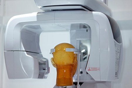
The study found that AI-based image processing may partly preserve image quality in low-dose cone-beam computed tomography (CBCT, a dental 3D imaging method) when the dose is moderately reduced. The 20% dose images processed with AI were not significantly different in quality from the 100% raw dose images, while the 10% dose images were worse.
Why the authors say this matters
The authors suggest this may help reduce the image-quality loss that comes with lowering CBCT radiation dose. They also conclude that larger studies in more diverse patient groups and clinical settings are needed to confirm the findings.
What the researchers tested
The researchers acquired CBCT scans from one healthy adult male at three dose levels: 10%, 20%, and 100% of standard dose. Each dataset was then processed with an AI-based image enhancement model, and five dental specialists rated the images using a 6-point scale across 12 anatomical and diagnostic criteria.
What worked and what didn't
AI-processed 20% dose images received image-quality scores that did not differ significantly from 100% raw dose images (median 4.45 vs. 5.05; p > 0.05). AI-processed 10% dose images scored significantly lower (p = 0.0074), and AI-processed 100% dose images were rated lower than the corresponding raw images.
What to keep in mind
This was a preliminary study based on scans from a single healthy adult male, so the results are limited in scope. The abstract also notes that further large-scale studies in diverse patient populations and clinical settings are required.
Key points
- AI image processing was tested as a way to improve low-dose dental CBCT image quality.
- At 20% of standard dose, AI-processed images were not significantly different from 100% raw dose images.
- At 10% of standard dose, AI-processed images had significantly lower quality scores.
- AI-processed 100% dose images were rated lower than the corresponding raw images.
- The study used scans from one healthy adult male and five dental specialists as raters.
Disclosure
- Research title:
- AI processing improved some low-dose CBCT images
- Authors:
- Tae-Yoon Park, Seung-Eun Lee, Sang-Yoon Park, Sung-Woon On, Sang-Min Yi, Byoung‐Eun Yang, Soo‐Hwan Byun
- Institutions:
- Hallym University, Hallym University, Hallym University, Hallym University, Hallym University, Hallym University, Hallym University Dongtan Sacred Heart Hospital, Hallym University Medical Center, Hallym University Medical Center, Hallym University Medical Center, Hallym University Medical Center, Hallym University Sacred Heart Hospital, Hallym University Sacred Heart Hospital, Hallym University Sacred Heart Hospital, Hallym University Sacred Heart Hospital, Hallym University Sacred Heart Hospital, Hallym University Sacred Heart Hospital
- Publication date:
- 2026-03-05
- OpenAlex record:
- View
- Image credit:
- FDA, Wikimedia Commons, Public domain
Get the weekly research newsletter
Stay current with scholarly research without reading academic papers — one filtered digest, every Friday.