What the study found
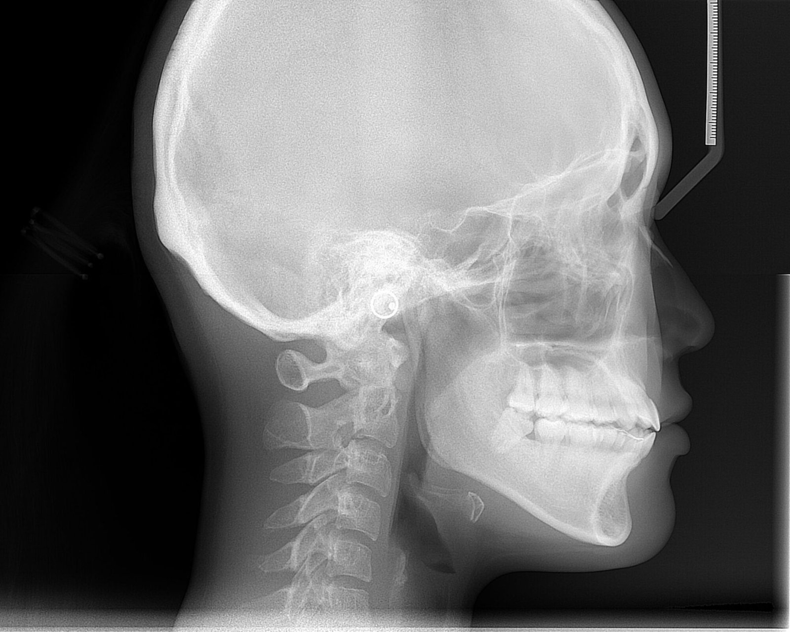
The study found that an end-to-end AI framework could identify and quantify age-related and sex-related patterns in craniofacial growth from lateral cephalometric radiographs, which are side-view X-ray images of the head. The authors report that the model visualized changing growth patterns across development and measured sex differences in craniofacial regions.
Why the authors say this matters
The authors conclude that the findings provide objective quantitative references for assessing developmental stages and for guiding the timing of interventions targeting specific craniofacial regions. They also say the results validate established developmental theories and offer new insights into coordinated craniofacial bone growth and sex-specific radiological characteristics.
What the researchers tested
The researchers developed an end-to-end deep learning framework using lateral cephalometric radiographs from 41,625 people aged 4 to 18 years. The model was designed to learn directly from the images without manual annotations, and Grad-CAM, or gradient-weighted class activation mapping, was used to create population-averaged saliency maps showing age-related and sex-related patterns. They also introduced two measures, the Age-related Saliency Index (ASI) and the Sex-related Saliency Index (SSI), to quantify the importance of developmental and sex-related features in craniofacial regions.
What worked and what didn't
Age-related saliency maps showed a shift in attention from external contours to internal bone details, and the ASI was used to prioritize these regions quantitatively. The SSI showed that sex differences were broadly distributed across cranial bones at earlier stages and became concentrated in the mandibular region by adulthood. The abstract does not describe failed analyses or negative results.
What to keep in mind
The summary provided does not describe specific limitations or external validation details. The findings are based on lateral cephalometric radiographs from ages 4 to 18, so the stated scope is limited to that developmental range and imaging type.
Key points
- An AI framework was trained on 41,625 lateral cephalometric radiographs from people aged 4 to 18 years.
- The model worked without manual annotations during training.
- Grad-CAM was used to show age-related and sex-related saliency patterns across craniofacial regions.
- Age-related attention shifted from external contours to internal bone details during development.
- Sex differences were widely distributed in early stages and became concentrated in the mandibular region by adulthood.
Disclosure
- Research title:
- Deep learning mapped craniofacial growth patterns across ages and sex
- Authors:
- Ziyi Hu, Yuyanran Zhang, Ningtao Liu, Xin Gao, Ziyu Huang, Guanglin Wu, Zhiyong Zhang, S W Wang
- Institutions:
- First Affiliated Hospital of Xi'an Jiaotong University, First Affiliated Hospital of Xi'an Jiaotong University, First Affiliated Hospital of Xi'an Jiaotong University, First Affiliated Hospital of Xi'an Jiaotong University, First Affiliated Hospital of Xi'an Jiaotong University, First Affiliated Hospital of Xi'an Jiaotong University, First Affiliated Hospital of Xi'an Jiaotong University, Luoyang Institute of Science and Technology, Second Affiliated Hospital of Xi'an Jiaotong University, Second Affiliated Hospital of Xi'an Jiaotong University, Second Affiliated Hospital of Xi'an Jiaotong University, Second Affiliated Hospital of Xi'an Jiaotong University, Second Affiliated Hospital of Xi'an Jiaotong University, Second Affiliated Hospital of Xi'an Jiaotong University, Second Affiliated Hospital of Xi'an Jiaotong University, Stomatology Hospital, Stomatology Hospital, Stomatology Hospital, Stomatology Hospital, Stomatology Hospital, Stomatology Hospital, Stomatology Hospital, Xi'an Jiaotong University, Xi'an Jiaotong University, Xi'an Jiaotong University, Xi'an Jiaotong University, Xi'an Jiaotong University, Xi'an Jiaotong University, Xi'an Jiaotong University
- Publication date:
- 2026-02-27
- OpenAlex record:
- View
- Image credit:
- ANUG, Wikimedia Commons, CC BY-SA 4.0
Get the weekly research newsletter
Stay current with scholarly research without reading academic papers — one filtered digest, every Friday.