What the study found
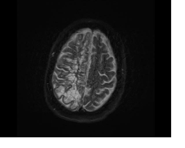
Higher MRI-visible perivascular spaces, or PVSs, in the basal ganglia were associated with worse cognitive performance over time. The association remained for executive function and visuospatial skills even after accounting for other cerebral small vessel disease markers.
Why the authors say this matters
The authors conclude that basal ganglia PVS burden may be an emerging marker of domain-specific cognitive decline in aging. They also say the findings support PVS as a vascular contributor to deep brain structure damage underlying cognitive decline over time.
What the researchers tested
PVS volumes were segmented with a deep learning algorithm. The researchers used linear mixed-effects models adjusted for age, sex, race/ethnicity, education, baseline cognitive status, apolipoprotein E-ε4 presence, Framingham Stroke Risk Profile, and intracranial volume, and they also ran head-to-head comparisons among statistically significant SVD markers.
What worked and what didn't
The abstract does not report the same independent result for the other cognitive measures after head-to-head comparison. The study also states that causation cannot be established.
What to keep in mind
This was an observational study, so it cannot show that PVS caused the cognitive decline. The abstract does not describe additional limitations beyond that point, and the findings come from one cohort of participants without stroke or dementia.
- Higher basal ganglia MRI-visible perivascular spaces were linked to worse cognitive performance over time.
- The strongest independent associations were with executive function and visuospatial skills.
- The study included 750 participants without stroke or dementia from a longitudinal cohort.
- MRI measures also included white matter hyperintensities, lacunes, and cerebral microbleeds.
- The authors say the findings support PVS as an emerging marker of domain-specific cognitive decline.